| |
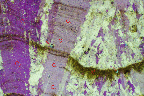
Figure 9-4. Columnar calcite (C), and a layer of microcrystalline CaCO3 (M) that ends abruptly at crystal C1 of the columnar calcite. Photomicrograph was taken in cross-polarized light with a gypsum plate in the light path, so that any one calcite crystal is all one shade of purple. Area labelled C1 is thus all one crystal. Red arrows point to quartz grains, one of which (at right) is in microcrystalline layer and other of which (at left) is in the columnar calcite but at a position laterally equivalent to the microcrystalline calcite. The abrupt termination of the layer of microcrystalline calcite against the columnar calcite and the presence of the quartz grain in the columnar calcite suggest that microcrystalline calcite extended leftward but has undergone recrystallization to form part of the columnar calcite crystals.
Another pair of
plane-light and
cross-polarized light images illustrate another example of evidence for recrystallization of microcrystalline CaCO3 to columnar calcite in this speleothem.
Photomicrograph was taken in cross-polarized light with a gypsum plate in the light path; A corresponding image shows the same area in plane-polarized light. Field of view is 2.3 mm wide. Carthage, Tennessee, U.S.A.; Stalagmite MR68; thin section MR68. Sample collected from highway construction debris by L. Bruce Railsback. |