Photomicrographs were taken in plane-polarized light. Anjohibe Cave, Madagascar; Stalagmite MA3; thin section MA3-18. Sample was collected by Dr. George A. Brook; celluluar identification was by Dr. John P. Shields.
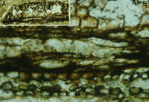
| Figure 6-5. Plant matter in a stalagmite from Madagascar. A (inset): Entire particle. Field of view is 1.2 mm wide. B (main panel): High-magnification view of part of grain in A.
The polygonal cells at the top are parenchyma that could be from a leaf or the cortex of a stem. The globular cells at the bottom are epidermis. Field of view is 0.21 mm. Photomicrographs were taken in plane-polarized light. Anjohibe Cave, Madagascar; Stalagmite MA3; thin section MA3-18. Sample was collected by Dr. George A. Brook; celluluar identification was by Dr. John P. Shields. |
|
|
|
| Back to the Table of Contents of the Atlas of Speleothem Microfabrics. |