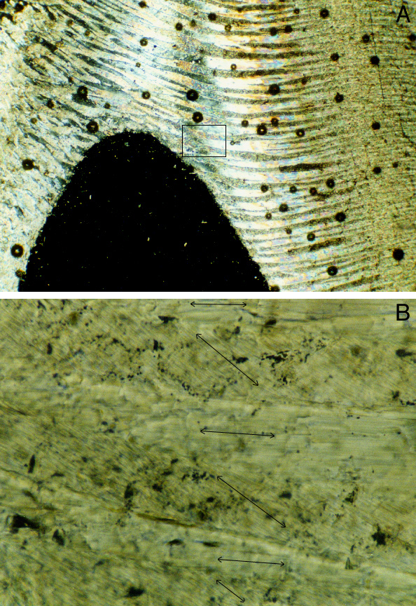
B: High-magnification image of area outlined in A. Image shows parts of six lamellae in A, and shows that each lamella consists of parallel aragonite crystals (black double-ended arrows are parallel to aragonite crystals). Sub-parallel lamellae in A thus consist of parallel aragonite crystals in B.
Photomicrograph A was taken in cross-polarized light; field of view is 2.2 mm wide. Photomicrograph B was taken in plane-polarized light; field of view is 0.21 mm wide. Modern shells; thin section GPODS. Sample collected by L. Bruce Railsback.