Figure 2-1. A: SEM image of a botryoid of aragonite in a stalagmite from Carlsbad Caverns. Aragonite (A) has nucleated on one very small CaCO3 grain (at arrow) and has grown over calcite (C). Scale bar is 100 microns. B: Higher magnification image of aragonite in the same botryoid. Scale bar is 10 microns.
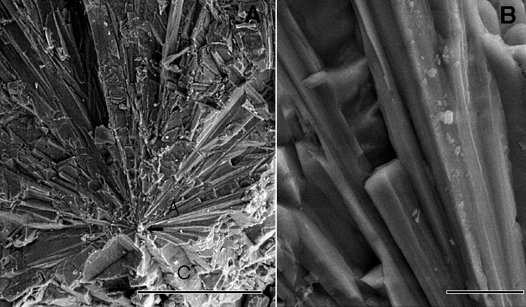
    Scanning electron microscope secondary electron images generated with 15 kV (A) and 20kV (B) acccelerating voltages. Georgia Giant speleothem, Carlsbad Caverns, New Mexico, U.S.A; Sample CB69. Sample collected by Dr. George A. Brook; image generated with the kind help of Dr. John P. Shields of the University of Georgia Center for Advanced Ultrastructural Research.
   
  Speleothem Image
   
   
  Back to the Table of Contents of the Atlas of Speleothem Microfabrics.