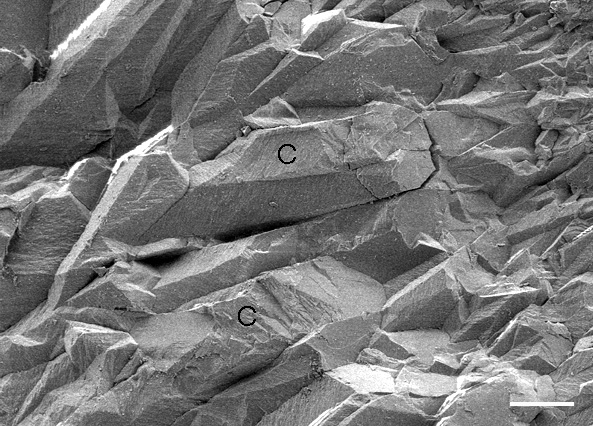
Scanning electron microscope secondary electron image generated with a 5 keV acccelerating voltage; white scale bar at lower right is 5 microns long. Georgia Giant speleothem, Carlsbad Caverns, New Mexico, U.S.A; Sample CB69. Sample collected by Dr. George A. Brook; image generated with the kind help of Dr. John P. Shields of the University of Georgia Center for Advanced Ultrastructural Research.