Scale bar at lower left is 10 microns long. Wonderwerk Cave, South Africa, Sample WW1, Mount WW1-2. Sample collected by Dr. George A. Brook; image generated with the kind help of Dr. John P. Shields.
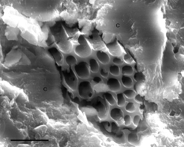
| Figure 6-7. Secondary electron SEM image of plant tissue (xylem) amidst calcite (C) in a stalagmite from South Africa. In a backscatter image, the plant fragment looks very dark compared to the surrounding calcite. An energy-dispersive X-ray spectrum of this material shows a large carbon peak, an oxygen peak, and a small calcium peak, whereas a spectrum from the surrounding calcite has a large Ca peak. The small Ca peak from the plant fragment presumably results from penetration of the SEM's electron beam through the fragment and into underlying calcite. Another image in this atlas shows a plant fragment lower in this stalagmite in transmitted light. Scale bar at lower left is 10 microns long. Wonderwerk Cave, South Africa, Sample WW1, Mount WW1-2. Sample collected by Dr. George A. Brook; image generated with the kind help of Dr. John P. Shields. |
|
|
|
| Back to the Table of Contents of the Atlas of Speleothem Microfabrics. |