Photomicrograph was taken in cross-polarized light with a gypsum plate in the light path; field of view is 0.39 mm wide. De Soto Caverns, Alabama, U.S.A.; Sample 95a; thin section 95a5. Sample collected by Dr. George A. Brook.
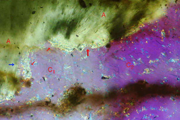
| Figure 9-9. High-magnification view of the base of an area of aragonite (A) shown in two lower-magnification images,
one in plane-polarized light and the other in cross-polarized light. C1 and C2 label calcite crystals shown in the image in cross-polarized light. Thick red arrow points to undercutting of aragonite, presumably as the result of dissolution and infilling by calcite. Thin red arrows point to edges of aragonite that was presumably a continuous botryoid and has undergone dissolution and replacement by calcite. Blue arrow points to calcite of crystal C1 that has engulfed remnants of aragonite marked by the lower of the two small diagonal red arrows. This evidence indicates that calcite has replaced aragonite, suggests that the aragonite shown in the lower-magnification images may have extended across the entire field of view of those images as one continuous layer, and invites speculation that all the calcite in those images may have originated in replacement of aragonite. Photomicrograph was taken in cross-polarized light with a gypsum plate in the light path; field of view is 0.39 mm wide. De Soto Caverns, Alabama, U.S.A.; Sample 95a; thin section 95a5. Sample collected by Dr. George A. Brook. |
|
|
|
| Back to the Table of Contents of the Atlas of Speleothem Microfabrics. |