Photomicrograph was taken in plane-polarized light; field of view is 2.3 mm wide. De Soto Caverns, Alabama, U.S.A.; Sample 95a; thin section 95a5. Sample collected by Dr. George A. Brook.
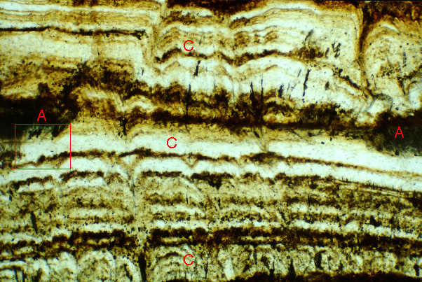
| Figure 9-7. Calcite (C) with isolated areas of aragonite (A) that lie along the same layer in a stalagmite. A corresponding image shows the same area in cross-polarized light, and a high-magnification image shows the area in the red rectangle. The high-magnification image presents evidence that calcite has replaced aragonite, suggesting that the aragonite may have extended across the entire field of view of this image as one continuous layer, and perhaps suggesting that all the calcite in this image may have originated in replacement of aragonite. Photomicrograph was taken in plane-polarized light; field of view is 2.3 mm wide. De Soto Caverns, Alabama, U.S.A.; Sample 95a; thin section 95a5. Sample collected by Dr. George A. Brook. |
|
|
|
| Back to the Table of Contents of the Atlas of Speleothem Microfabrics. |